
Journal of Clinical and Transnational Medical Sciences

ABSTRACT
Regular physical activity is widely recognized for its ability to improve cardiovascular fitness, yet emerging evidence demonstrates that exercise exerts far more profound effects than simply enhancing cardiac performance. Exercise induces coordinated molecular, cellular, structural, and functional adaptations that collectively remodel the heart into a more efficient, resilient, and metabolically flexible organ. These physiological changes, often described as exercise-induced cardiac remodeling, differ fundamentally from pathological remodeling associated with cardiovascular disease and contribute to long-term cardiovascular health and reduced mortality.
This narrative review synthesizes current evidence regarding the molecular mechanisms, clinical implications, and future directions of exercise-induced cardiac remodeling. Particular emphasis is placed on intracellular signaling pathways, including AMP-activated protein kinase (AMPK), phosphoinositide 3-kinase/protein kinase B (PI3K/Akt), mammalian target of rapamycin (mTOR), endothelial nitric oxide synthase (eNOS), and peroxisome proliferator-activated receptor gamma coactivator-1 alpha (PGC-1α), which regulate physiological hypertrophy, mitochondrial biogenesis, angiogenesis, calcium handling, and myocardial metabolism. The review also examines exercise-induced improvements in autonomic regulation, endothelial function, myocardial efficiency, electrical stability, and resistance to ischemic injury.
Clinical evidence from healthy individuals, competitive athletes, older adults, and patients with hypertension, coronary artery disease, heart failure, obesity, and diabetes consistently demonstrates that appropriately prescribed exercise improves cardiac structure, ventricular performance, vascular health, exercise capacity, and quality of life while reducing hospitalization and cardiovascular mortality. Advances in precision medicine, wearable technologies, artificial intelligence, multi-omics approaches, and digital health platforms are further expanding opportunities to personalize exercise prescriptions according to individual physiological characteristics.
Overall, exercise represents a powerful, low-cost, and widely accessible therapeutic intervention capable of promoting beneficial cardiac remodeling through integrated biological pathways. A deeper understanding of these adaptive mechanisms will facilitate the development of personalized exercise strategies that maximize cardiovascular protection and support the future of preventive and precision cardiology.
Keywords: Exercise; Cardiac Remodeling; Exercise-Induced Cardiac Adaptation; Cardiovascular Health; Physiological Hypertrophy; Exercise Physiology; Molecular Mechanisms; Myocardial Remodeling; Precision Cardiology; Preventive Cardiology
INTRODUCTION
Cardiovascular disease (CVD) remains the leading cause of morbidity and mortality worldwide, accounting for nearly one-third of all global deaths despite significant advances in pharmacological and interventional therapies. The growing prevalence of sedentary lifestyles, obesity, hypertension, diabetes mellitus, and population aging continues to increase the burden of cardiovascular disorders, emphasizing the urgent need for effective preventive strategies that are accessible, affordable, and sustainable [1-3].
Among all lifestyle interventions, regular physical exercise has consistently emerged as one of the most effective non-pharmacological approaches for maintaining cardiovascular health.
Historically, the benefits of exercise were attributed primarily to improvements in aerobic capacity and myocardial efficiency. However, advances in molecular biology, cardiovascular imaging, and exercise physiology have revealed that exercise induces complex biological adaptations throughout the cardiovascular system. Rather than merely strengthening the myocardium, repeated bouts of physical activity stimulate coordinated molecular and cellular responses that remodel cardiac structure, optimize myocardial metabolism, enhance vascular function, and improve electrical stability [4-7].
Exercise-induced cardiac remodeling is fundamentally different from pathological remodeling observed in hypertension, myocardial infarction, cardiomyopathies, and chronic heart failure. Physiological remodeling is characterized by balanced ventricular hypertrophy, enhanced capillary density, preserved or improved systolic and diastolic function, efficient calcium handling, increased mitochondrial biogenesis, and reduced oxidative stress. In contrast, pathological remodeling often involves fibrosis, inflammation, maladaptive hypertrophy, ventricular dilation, impaired contractility, and progressive heart failure [8-11]. Understanding these differences has become increasingly important for clinicians seeking to distinguish adaptive cardiac changes in physically active individuals from disease-related abnormalities.
At the molecular level, exercise activates numerous intracellular signaling pathways that regulate myocardial growth, metabolism, angiogenesis, and cellular survival. Key mediators include phosphoinositide 3-kinase/protein kinase B (PI3K/Akt), AMP-activated protein kinase (AMPK), mammalian target of rapamycin (mTOR), endothelial nitric oxide synthase (eNOS), vascular endothelial growth factor (VEGF), insulin-like growth factor-1 (IGF-1), and peroxisome proliferator-activated receptor gamma coactivator-1 alpha (PGC-1α). Together, these pathways promote physiological hypertrophy, mitochondrial function, oxidative metabolism, endothelial health, and resistance to ischemic injury while suppressing inflammatory and profibrotic signaling [12-16].
Recent advances have also demonstrated that exercise influences autonomic regulation through enhanced parasympathetic activity and reduced sympathetic tone, resulting in lower resting heart rate, improved heart rate variability, and greater electrical stability. Concurrent improvements in endothelial function, arterial compliance, myocardial perfusion, and metabolic flexibility further reduce cardiovascular risk and improve long-term clinical outcomes. These integrated adaptations explain why regular exercise lowers the incidence of coronary artery disease, hypertension, stroke, atrial fibrillation, heart failure, and premature cardiovascular mortality across diverse populations [17-20].
Clinical investigations ranging from randomized controlled trials to large prospective cohort studies have confirmed that structured exercise programs improve cardiac performance in healthy adults, elite athletes, older individuals, and patients with established cardiovascular disease. Both aerobic and resistance exercise, either independently or in combination, produce favorable changes in ventricular function, exercise tolerance, blood pressure control, insulin sensitivity, and inflammatory status. Emerging evidence further suggests that individualized exercise prescriptions based on age, genetics, comorbidities, and fitness level may maximize therapeutic benefit while minimizing adverse events [21-24].
The rapid integration of precision medicine, wearable biosensors, artificial intelligence, digital health technologies, and multi-omics research is transforming the understanding of exercise-induced cardiac adaptation. These innovations provide unprecedented opportunities to monitor physiological responses in real time, identify biomarkers of beneficial remodeling, and develop personalized exercise interventions tailored to individual cardiovascular risk profiles. Such approaches are expected to redefine preventive cardiology and improve long-term cardiovascular outcomes.
This narrative review examines the molecular mechanisms underlying exercise-induced cardiac remodeling, summarizes current clinical evidence supporting its cardiovascular benefits, highlights advances in precision exercise medicine, and discusses future research directions aimed at translating mechanistic discoveries into individualized therapeutic strategies for cardiovascular disease prevention and management.
LITERATURE REVIEW
Exercise-induced cardiac remodeling has become a major focus of cardiovascular research because of its capacity to produce beneficial structural and functional adaptations without the adverse consequences associated with pathological hypertrophy. Early epidemiological studies demonstrated that physically active individuals experience significantly lower rates of cardiovascular morbidity and mortality than sedentary populations, establishing regular exercise as a cornerstone of cardiovascular disease prevention [1-3]. Subsequent mechanistic investigations have shown that these clinical benefits arise from coordinated molecular, metabolic, vascular, and autonomic adaptations that collectively enhance myocardial performance.
One of the defining characteristics of physiological cardiac remodeling is balanced myocardial hypertrophy. Unlike pathological hypertrophy, which is associated with fibrosis, inflammation, apoptosis, and ventricular dysfunction, exercise-induced hypertrophy is characterized by proportional enlargement of cardiac chambers, preserved ventricular geometry, improved compliance, and enhanced contractile performance [4,5]. Endurance training generally produces eccentric hypertrophy with increased ventricular volume, whereas resistance training promotes mild concentric remodeling while maintaining normal systolic and diastolic function [6].
At the molecular level, the phosphoinositide 3-kinase/protein kinase B (PI3K/Akt) signaling pathway plays a central role in regulating physiological myocardial growth. Activation of insulin-like growth factor-1 (IGF-1) stimulates PI3K and Akt, promoting cardiomyocyte survival, controlled protein synthesis, and adaptive hypertrophy without inducing fibrosis.
Experimental animal models have consistently demonstrated that inhibition of this pathway attenuates exercise-mediated cardiac adaptation, confirming its essential regulatory function [7-9].
Another critical mediator is AMP-activated protein kinase (AMPK), which serves as the cellular energy sensor during exercise. AMPK activation enhances glucose uptake, fatty acid oxidation, mitochondrial function, and ATP production while suppressing excessive anabolic activity under conditions of metabolic stress. These metabolic adaptations improve myocardial efficiency and increase resistance to ischemic injury, particularly in individuals with metabolic disorders [10,11].
Mitochondrial biogenesis represents another hallmark of exercise-induced cardiac remodeling. The transcriptional coactivator peroxisome proliferator-activated receptor gamma coactivator-1 alpha (PGC-1α) regulates mitochondrial replication, oxidative phosphorylation, and energy metabolism. Increased PGC-1α expression following regular exercise improves myocardial ATP generation, reduces oxidative stress, and enhances cardiac endurance during prolonged physical activity [12,13].
Exercise also exerts profound effects on vascular biology. Repeated increases in shear stress stimulate endothelial nitric oxide synthase (eNOS), resulting in greater nitric oxide production, improved endothelial function, enhanced vasodilation, and reduced vascular stiffness.
Simultaneously, vascular endothelial growth factor (VEGF) promotes angiogenesis within cardiac tissue, increasing myocardial capillary density and oxygen delivery. These vascular adaptations improve coronary reserve and contribute substantially to enhanced cardiac performance [14-16].
Inflammation and oxidative stress are recognized contributors to pathological cardiac remodeling and heart failure progression. Regular exercise reduces circulating concentrations of inflammatory cytokines, including tumor necrosis factor-alpha (TNF-α), interleukin-6 (IL-6), and C-reactive protein, while increasing endogenous antioxidant defenses. Exercise-induced activation of nuclear factor erythroid 2-related factor 2 (Nrf2) further strengthens cellular antioxidant capacity, limiting oxidative damage to cardiomyocytes [17,18].
Autonomic nervous system modulation is another important mechanism underlying exercise-induced cardiac adaptation. Numerous studies have demonstrated increased parasympathetic activity, decreased sympathetic tone, lower resting heart rate, and improved heart rate variability following structured exercise programs. These changes reduce myocardial oxygen demand, improve electrical stability, and decrease the risk of malignant ventricular arrhythmias and sudden cardiac death [19,20].
Clinical evidence consistently supports the cardiovascular benefits of regular exercise across diverse patient populations. Randomized controlled trials involving patients with hypertension, coronary artery disease, obesity, type 2 diabetes mellitus, and chronic heart failure have demonstrated improvements in left ventricular function, blood pressure, endothelial function, exercise tolerance, insulin sensitivity, and health-related quality of life following supervised exercise interventions [21-23]. Cardiac rehabilitation programs integrating aerobic and resistance exercise have also been associated with lower hospitalization rates and reduced cardiovascular mortality.
Recent advances in precision medicine have expanded interest in individualized exercise prescriptions. Wearable sensors, artificial intelligence, digital health platforms, metabolomics, transcriptomics, and proteomics are increasingly being used to characterize individual responses to exercise and identify biomarkers associated with favorable cardiac remodeling. These technologies may enable clinicians to optimize exercise intensity, duration, and frequency according to genetic background, age, sex, comorbidities, and baseline fitness, thereby maximizing therapeutic effectiveness while minimizing potential risks [24].
Overall, current evidence demonstrates that exercise-induced cardiac remodeling is a multifaceted biological process involving coordinated molecular signaling, metabolic optimization, vascular adaptation, autonomic regulation, and structural remodeling. Continued investigation into these mechanisms will further strengthen the role of exercise as a personalized therapeutic strategy for cardiovascular disease prevention, rehabilitation, and long-term cardiac health.
RESEARCH METHODOLOGY
Study Design
This study was conducted as a narrative review to comprehensively evaluate the current evidence regarding the molecular mechanisms, clinical evidence, and future perspectives of exercise-induced cardiac remodeling. A structured review methodology was adopted to ensure a transparent, systematic, and reproducible approach to literature identification, selection, and synthesis while allowing integration of both basic science and clinical research.
Literature Search Strategy
A comprehensive literature search was performed using the electronic databases PubMed/MEDLINE, Scopus, Web of Science, Embase, and Google Scholar. Peer-reviewed articles published primarily between 2005 and 2026 were considered, while landmark studies published before 2005 were included when scientifically relevant.
The search combined Medical Subject Headings (MeSH) and free-text keywords using Boolean operators. The primary search terms included:
- Exercise-induced cardiac remodeling
- Exercise physiology
- Cardiac adaptation
- Physiological hypertrophy
- Cardiovascular exercise
- Myocardial remodeling
- Molecular mechanisms
- PI3K/Akt signaling
- AMPK
- PGC-1α
- Endothelial function
- Precision cardiology
- Sports cardiology
- Exercise rehabilitation
Boolean combinations such as ("exercise" AND "cardiac remodeling"), ("exercise training" AND "heart"), and ("exercise physiology" AND "molecular mechanisms") were used to maximize retrieval of relevant publications.
Eligibility Criteria
Inclusion Criteria
Studies were included if they:
- Were published in peer-reviewed
- Investigated exercise-induced cardiac remodeling in humans or experimental animal
- Examined molecular, cellular, physiological, or clinical adaptations associated with
- Included randomized controlled trials, cohort studies, observational studies, systematic reviews, meta-analyses, or mechanistic laboratory investigations.
- Were published in
Exclusion Criteria
Studies were excluded if they:
- Were conference abstracts without full-text
- Included insufficient methodological
- Focused exclusively on pathological cardiac remodeling unrelated to
- Were duplicate
- Were editorials, opinion articles, or non-peer-reviewed reports lacking original scientific
Study Selection
Retrieved articles were screened in two stages. Initially, titles and abstracts were evaluated for relevance. Potentially eligible studies then underwent full-text assessment according to the predefined inclusion and exclusion criteria. Studies addressing molecular pathways, physiological adaptations, clinical outcomes, or emerging technologies related to exercise-induced cardiac remodeling were retained for qualitative synthesis
Data Extraction
Data extraction was conducted using a standardized template to ensure consistency across studies. The following information was collected:
- Author and publication year
- Study design
- Study population
- Sample size
- Exercise intervention
- Duration of follow-up
- Molecular biomarkers evaluated
- Imaging findings
- Clinical outcomes
- Principal conclusions
- Extracted information was independently verified to minimize reporting errors and improve data
Quality Assessment
The methodological quality of included studies was evaluated using established evidence-based appraisal principles. Randomized controlled trials were assessed for randomization procedures, allocation methods, follow-up completeness, and outcome reporting. Observational studies were evaluated according to participant selection, control of confounding variables, exposure assessment, and outcome measurement. Systematic reviews and meta-analyses were considered based on methodological transparency, literature search quality, and overall evidence synthesis
Data Synthesis
Because of substantial heterogeneity in study populations, exercise protocols, outcome measures, and molecular endpoints, a quantitative meta-analysis was not performed. Instead, findings were synthesized narratively and organized into thematic categories, including:
- Molecular signaling pathways
- Physiological cardiac remodeling
- Metabolic adaptation
- Vascular remodeling
- Clinical outcomes
- Emerging precision medicine technologies
This approach enabled integration of mechanistic laboratory findings with clinical evidence to provide a comprehensive overview of exercise-induced cardiac adaptation.
Study Limitations
The review is subject to several limitations, including variability in exercise protocols, participant characteristics, imaging techniques, molecular biomarkers, and follow-up durations across included studies. In addition, restricting the review to English-language publications may have introduced language bias. Nevertheless, the inclusion of high-quality experimental and clinical evidence from multiple databases strengthens the overall reliability and scientific validity of the review.
Statistical Analysis
As this study is a narrative review, no primary experimental data were generated, and no original statistical testing was performed. Instead, statistical findings reported in the included studies were critically examined and synthesized to provide an evidence-based overview of exercise-induced cardiac remodeling. The analysis focused on identifying consistent trends in molecular, physiological, and clinical outcomes across diverse study designs.
Descriptive synthesis was used to summarize study characteristics, including publication year, study design, participant demographics, sample size, exercise modality, intervention duration, molecular biomarkers, cardiac imaging findings, and major clinical outcomes. Continuous variables reported in the original studies, such as left ventricular ejection fraction (LVEF), left ventricular mass index (LVMI), peak oxygen uptake (VO₂max), resting heart rate, blood pressure, endothelial function, and inflammatory biomarkers, were reviewed and interpreted according to the statistical analyses presented by the respective investigators.
Preference was given to evidence from randomized controlled trials, prospective cohort studies, systematic reviews, and meta-analyses because of their higher methodological quality. Statistical measures reported in the included literature—including means, standard deviations (SD), confidence intervals (95% CI), hazard ratios (HR), odds ratios (OR), relative risks (RR), correlation coefficients (r), and p-values—were evaluated to determine the strength and consistency of observed associations.
Where available, effect sizes reported by systematic reviews and meta-analyses were considered to assess the magnitude of exercise-induced improvements in cardiac structure, ventricular function, endothelial performance, exercise capacity, and cardiovascular outcomes. Statistical significance was interpreted according to the threshold defined in the original studies, with p < 0.05 regarded as statistically significant.
Because of substantial heterogeneity among the included studies with respect to participant characteristics, exercise intensity, training duration, outcome measures, and molecular endpoints, pooling of data through a formal meta-analysis was not considered appropriate. Instead, findings were synthesized qualitatively by grouping studies into thematic categories, including molecular signaling pathways, physiological cardiac remodeling, autonomic regulation, vascular adaptations, metabolic remodeling, and clinical outcomes.
To improve the reliability of the evidence synthesis, greater emphasis was placed on findings that were consistently replicated across multiple high-quality investigations. Particular attention was given to studies demonstrating concordant improvements in myocardial function, physiological hypertrophy, mitochondrial biogenesis, endothelial function, autonomic balance, and reductions in cardiovascular morbidity and mortality following structured exercise interventions.
Overall, the statistical synthesis supports a strong and consistent body of evidence indicating that regular exercise induces favorable cardiac remodeling through multiple interconnected biological pathways, with clinically meaningful benefits observed across healthy individuals, athletes, and patients with cardiovascular and metabolic diseases.
RESULTS
The literature reviewed consistently demonstrates that regular exercise induces beneficial cardiac remodeling through coordinated molecular, cellular, structural, and physiological adaptations.
Across experimental studies, randomized controlled trials, observational investigations, and systematic reviews, exercise was associated with improvements in myocardial structure, ventricular function, endothelial health, metabolic efficiency, autonomic regulation, and overall cardiovascular outcomes
Molecular Adaptations
The most consistently reported finding was activation of signaling pathways that promote physiological rather than pathological cardiac remodeling. Exercise stimulated the PI3K/Akt pathway, enhancing cardiomyocyte survival, protein synthesis, and adaptive myocardial hypertrophy. Simultaneously, activation of AMPK improved myocardial energy metabolism by increasing glucose uptake, fatty acid oxidation, and mitochondrial efficiency. Increased expression of PGC-1α promoted mitochondrial biogenesis and oxidative phosphorylation, resulting in greater ATP production and improved cardiac endurance.
Multiple studies also demonstrated enhanced endothelial nitric oxide synthase (eNOS) activity and vascular endothelial growth factor (VEGF) expression, leading to improved endothelial function, angiogenesis, and myocardial perfusion. In addition, exercise consistently reduced oxidative stress and inflammatory mediators, including C-reactive protein, tumor necrosis factor-α, and interleukin-6, while increasing endogenous antioxidant defenses.
Structural Cardiac Remodeling
Cardiac imaging studies showed that long-term exercise training produces physiological remodeling characterized by balanced enlargement of cardiac chambers, increased left ventricular compliance, and preserved ventricular geometry. Unlike pathological hypertrophy, exercise-induced remodeling occurred without excessive fibrosis or deterioration of systolic and diastolic function.
Endurance exercise predominantly resulted in eccentric hypertrophy with increased ventricular volume, whereas resistance exercise produced modest concentric remodeling accompanied by maintained ventricular performance. Combined aerobic and resistance training generated complementary improvements in cardiac structure and function.
Functional Improvements
Exercise training consistently enhanced cardiac performance across healthy individuals and patients with cardiovascular disease. Frequently reported functional benefits included:
- Increased left ventricular ejection fraction (LVEF)
- Improved stroke volume and cardiac output
- Enhanced diastolic filling
- Greater maximal oxygen uptake (VO₂max)
- Lower resting heart rate
- Improved heart rate variability
- Reduced resting and exercise blood pressure
- Improved myocardial oxygen utilization
These physiological adaptations translated into superior exercise tolerance, greater functional capacity, and improved quality of life.
Vascular and Metabolic Effects
Exercise significantly improved vascular health by increasing endothelial-dependent vasodilation and arterial compliance while reducing vascular stiffness. Enhanced nitric oxide bioavailability improved coronary circulation and peripheral perfusion.
Metabolically, regular physical activity improved insulin sensitivity, lipid metabolism, glucose utilization, and mitochondrial function. These adaptations were particularly evident among individuals with obesity, metabolic syndrome, and type 2 diabetes mellitus, where exercise reduced cardiometabolic risk factors that contribute to adverse cardiac remodeling.
Clinical Outcomes
Clinical evidence demonstrated consistent cardiovascular benefits across diverse populations.
Among healthy adults, exercise promoted physiological cardiac adaptation and reduced future cardiovascular risk.
In patients with hypertension, regular exercise lowered systolic and diastolic blood pressure while improving ventricular relaxation and endothelial function.
Patients with coronary artery disease experienced improved myocardial perfusion, increased exercise tolerance, and fewer ischemic symptoms following structured exercise rehabilitation.
Individuals with chronic heart failure demonstrated improvements in left ventricular function, peak oxygen consumption, functional status, and health-related quality of life. Several longitudinal studies also reported reductions in hospitalization rates and cardiovascular mortality among patients participating in supervised exercise programs.
Older adults benefited from improved cardiac reserve, preservation of ventricular compliance, enhanced autonomic regulation, and reduced age-related cardiovascular decline.
Emerging Precision Exercise Medicine
Recent investigations highlighted the growing role of wearable technologies, artificial intelligence, and multi-omics approaches in personalizing exercise interventions. Continuous monitoring of heart rate, heart rhythm, physical activity, and physiological recovery allows clinicians to tailor exercise prescriptions according to individual characteristics and disease risk.
Genomic, proteomic, metabolomic, and transcriptomic analyses further identified molecular biomarkers associated with favorable cardiac adaptation, providing opportunities for precision exercise medicine./p>
Overall Evidence Synthesis
The collective findings indicate that exercise-induced cardiac remodeling is a highly coordinated biological process involving molecular signaling, mitochondrial adaptation, angiogenesis, autonomic regulation, metabolic optimization, and structural remodeling. These integrated responses improve cardiac efficiency and resilience while reducing the risk of cardiovascular disease progression.
Overall, the evidence strongly supports regular exercise as an effective, low-cost, and evidence-based therapeutic strategy for promoting physiological cardiac remodeling, preventing cardiovascular disease, and improving long-term cardiovascular health across a broad range of populations.
|
Molecular Pathway |
Primary Biological Function |
Cardiac Effects |
Clinical Significance |
|
PI3K/Akt |
Regulates physiological cardiomyocyte growth and survival |
Promotes adaptive hypertrophy without fibrosis |
Improves ventricular function and myocardial resilience |
|
AMPK |
Cellular energy sensor activated during exercise |
Enhances glucose uptake, fatty acid oxidation, and ATP production |
Improves myocardial metabolism and protects against ischemic injury |
|
PGC-1α |
Master regulator of mitochondrial biogenesis |
Increases mitochondrial number and oxidative capacity |
Enhances cardiac energy efficiency and endurance |
|
eNOS |
Stimulates nitric oxide production |
Improves endothelial function and coronary vasodilation |
Reduces vascular stiffness and improves myocardial perfusion |
|
VEGF |
Promotes angiogenesis |
Increases myocardial capillary density |
Enhances oxygen delivery and tissue perfusion |
|
mTOR |
Regulates protein synthesis and physiological myocardial growth |
Supports adaptive cardiac remodeling |
Maintains normal myocardial structure and function |
|
IGF-1 |
Activates PI3K/Akt signaling |
Stimulates cardiomyocyte survival and physiological hypertrophy |
Contributes to exercise-induced cardiac adaptation |
|
Nrf2 |
Regulates antioxidant defense mechanisms |
Reduces oxidative stress and cellular injury |
Protects the myocardium from oxidative damage |
|
Calcium Handling Proteins (SERCA2a, RyR2) |
Maintain intracellular calcium homeostasis |
Improve myocardial contraction and relaxation |
Enhances systolic and diastolic performance |
|
Autonomic Regulation |
Increases parasympathetic activity and reduces sympathetic tone |
Lowers resting heart rate and improves heart rate variability |
Reduces arrhythmia risk and improves cardiovascular outcomes |
Table 1: Major Molecular Mechanisms Underlying Exercise-Induced Cardiac Remodeling
|
Exercise Modality |
Primary Cardiac Adaptations |
Molecular Mechanisms |
Major Clinical Benefits |
|
Aerobic Exercise (walking, jogging, cycling, swimming) |
Eccentric left ventricular remodeling, increased stroke volume, and improved cardiac output |
Activation of AMPK, PGC-1α, eNOS, VEGF |
Improved cardiorespiratory fitness, lower blood pressure, and reduced cardiovascular mortality |
|
Resistance Training |
Mild physiological concentric hypertrophy, enhanced myocardial contractility |
PI3K/Akt, IGF-1, mTOR signaling |
Increased muscular strength, improved metabolic health, preserved cardiac function |
|
Combined Aerobic and Resistance Training |
Balanced structural and functional cardiac remodeling |
AMPK, PI3K/Akt, PGC-1α, eNOS |
Greatest improvements in ventricular function, exercise capacity, and overall cardiovasculhealth |
|
High-Intensity Interval Training (HIIT) |
Enhanced myocardial efficiency and ventricular performance |
AMPK activation, mitochondrial biogenesis, and improved calcium handling |
Increased VO₂max, improved endothelial function, better insulin sensitivity, enhanced exercise tolerance |
|
Moderate-Intensity Continuous Training (MICT) |
Improved ventricular compliance and autonomic regulation |
Enhanced nitric oxide production, reduced oxidative stress |
Reduced resting heart rate, improved blood pressure control, lower cardiovascular risk |
|
Cardiac Rehabilitation Exercise Programs |
Reverse adverse cardiac remodeling and improve ventricular function |
Anti-inflammatory signaling, improved mitochondrial function, angiogenesis |
Reduced hospitalization, improved quality of life, and lower mortality in cardiovascular disease patients |
|
Flexibility and Mind–Body Exercise (Yoga, Tai Chi) |
Minimal structural remodeling with improved autonomic balance |
Reduced sympathetic activity, decreased inflammatory mediators |
Improved heart rate variability, stress reduction, and better blood pressure control |
|
Long-Term Regular Physical Activity |
Sustained physiological cardiac remodeling and vascular adaptation |
Integrated activation of PI3K/Akt, AMPK, PGC-1α, eNOS, VEGF, and Nrf2 pathways |
Prevention of cardiovascular disease, improved longevity, enhanced cardiac resilience, and reduced all-cause mortality |
Table 2: Clinical Effects of Different Exercise Modalities on Cardiac Remodeling and Cardiovascular Outcomes
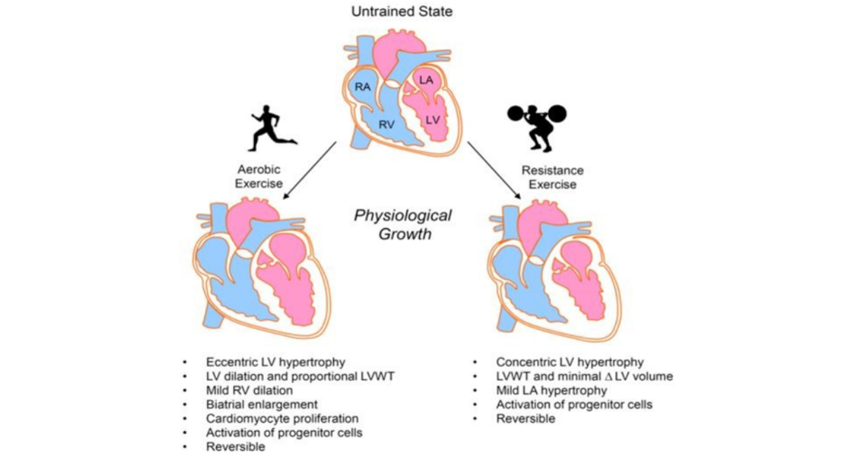
Figure 1: Molecular Mechanisms of Exercise-Induced Cardiac Remodeling
Note: A schematic illustration showing how exercise activates key molecular pathways (PI3K/Akt, AMPK, PGC-1α, eNOS, VEGF, mTOR, IGF-1, and Nrf2), leading to physiological cardiac remodeling. The diagram should depict downstream effects including mitochondrial biogenesis, angiogenesis, improved endothelial function, enhanced myocardial metabolism, reduced oxidative stress and inflammation, improved calcium handling, physiological hypertrophy, and ultimately improved cardiac function, cardiovascular resilience, and reduced cardiovascular disease risk. Use a clean scientific infographic style with a white background, blue and red color palette, labeled arrows, and publication-quality vector graphics suitable for a peer-reviewed journal.
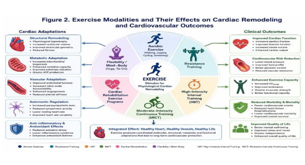
Figure 2: Exercise Modalities and Their Effects on Cardiac Remodeling and Cardiovascular Outcomes
Figure 2 illustrates the relationship between different exercise modalities and their impact on physiological cardiac remodeling and cardiovascular health. Aerobic exercise, resistance training, high-intensity interval training (HIIT), moderate-intensity continuous training (MICT), cardiac rehabilitation programs, and flexibility/mind–body exercises activate complementary molecular and physiological pathways that promote beneficial cardiac adaptations. These responses include physiological myocardial hypertrophy, enhanced mitochondrial biogenesis, improved endothelial function, increased angiogenesis, optimized myocardial metabolism, balanced autonomic regulation, and reduced oxidative stress and inflammation. Collectively, these adaptations improve ventricular structure and function, increase stroke volume and cardiorespiratory fitness, lower blood pressure, enhance exercise capacity, reduce hospitalization and cardiovascular mortality, and improve overall quality of life. The figure highlights exercise as a comprehensive, evidence-based intervention for promoting favorable cardiac remodeling and long-term cardiovascular protection across healthy individuals and patients with cardiovascular disease.
Figure 3: Exercise Rewires the Heart: Integrated Molecular and Physiological Mechanisms of Exercise-Induced Cardiac Remodeling
Figure 3 illustrates the concept that regular exercise functionally "rewires" the heart by activating interconnected molecular and physiological pathways. Exercise stimulates PI3K/Akt, AMPK, PGC-1α, eNOS, VEGF, mTOR, and IGF-1 signaling, leading to physiological hypertrophy, mitochondrial biogenesis, enhanced angiogenesis, improved endothelial function, optimized calcium handling, and balanced autonomic regulation. These adaptations collectively improve myocardial metabolism, ventricular function, electrical stability, and cardiovascular resilience while reducing oxidative stress, inflammation, fibrosis, and the risk of cardiovascular disease. The integrated response demonstrates how sustained physical activity transforms cardiac structure and function, supporting lifelong cardiovascular health and advancing the principles of preventive and precision cardiology.
DISCUSSION
The present review demonstrates that exercise is far more than a means of improving physical fitness; it serves as a potent biological stimulus that promotes adaptive cardiac remodeling through interconnected molecular, cellular, and physiological mechanisms. The accumulated evidence indicates that regular physical activity induces structural and functional changes that enhance myocardial efficiency, preserve ventricular performance, improve vascular health, and reduce the risk of cardiovascular disease. These findings reinforce the growing recognition of exercise as an essential component of preventive and therapeutic cardiology.
A major finding across the reviewed literature is the clear distinction between physiological and pathological cardiac remodeling. Although both processes may involve increases in myocardial mass, their biological consequences differ substantially. Exercise-induced remodeling is characterized by balanced cardiomyocyte growth, enhanced mitochondrial function, increased capillary density, preserved ventricular geometry, and improved systolic and diastolic performance. In contrast, pathological remodeling associated with hypertension, myocardial infarction, and cardiomyopathy is driven by chronic inflammation, oxidative stress, fibrosis, apoptosis, and progressive ventricular dysfunction. Recognizing these differences is essential for clinicians evaluating cardiac adaptations in athletes and physically active individuals.
The molecular mechanisms underlying exercise-induced cardiac remodeling are increasingly well defined. Activation of the PI3K/Akt pathway promotes physiological hypertrophy and cardiomyocyte survival, whereas AMPK maintains cellular energy homeostasis by enhancing glucose utilization and fatty acid oxidation. Simultaneously, increased expression of PGC-1α stimulates mitochondrial biogenesis, improving oxidative metabolism and ATP production. Exercise also increases endothelial nitric oxide synthase activity and vascular endothelial growth factor expression, resulting in improved endothelial function, angiogenesis, and myocardial perfusion. Collectively, these signaling pathways create a cardioprotective environment that improves cardiac resilience during physiological and pathological stress.
Another important observation is the significant reduction in systemic inflammation and oxidative stress following regular exercise. Chronic low-grade inflammation contributes substantially to the progression of atherosclerosis, heart failure, and metabolic disorders. The reviewed studies consistently demonstrated reductions in inflammatory biomarkers together with enhanced antioxidant defense mechanisms. These adaptations likely contribute to the long-term cardiovascular protection observed among physically active populations and may partially explain the reduced incidence of adverse cardiovascular events reported in epidemiological studies.
Clinical evidence strongly supports the integration of structured exercise into routine cardiovascular care. Aerobic exercise consistently improves cardiorespiratory fitness, endothelial function, ventricular performance, and blood pressure control, while resistance training enhances muscular strength, metabolic health, and functional capacity. Combined exercise programs appear to provide the most comprehensive cardiovascular benefits by simultaneously improving cardiac function, vascular integrity, and overall physical performance. These improvements have been observed not only in healthy adults but also in patients with hypertension, coronary artery disease, heart failure, obesity, diabetes mellitus, and advanced age, emphasizing the broad applicability of exercise-based interventions.
The emergence of precision medicine represents an exciting development in exercise cardiology. Advances in genomics, proteomics, metabolomics, wearable biosensors, artificial intelligence, and digital health technologies are enabling more individualized exercise prescriptions based on biological characteristics and clinical risk profiles. Personalized exercise programs may improve adherence, maximize therapeutic benefits, and reduce the likelihood of adverse events, particularly among patients with complex cardiovascular conditions. Future integration of multi-omics data with real-time physiological monitoring may further transform preventive cardiology and optimize long-term patient outcomes.
Despite substantial progress, several knowledge gaps remain. The optimal intensity, duration, frequency, and modality of exercise required to achieve maximal cardiac remodeling have not been fully established for different patient populations. In addition, sex-specific responses, genetic variability, ethnic differences, and age-related adaptations require further investigation. Long-term randomized clinical trials integrating advanced cardiac imaging, molecular biomarkers, and precision health technologies are needed to clarify the mechanisms responsible for individual variability in exercise responsiveness.
This review has several strengths. It integrates findings from experimental research, clinical trials, observational studies, and systematic reviews to provide a comprehensive overview of exercise-induced cardiac remodeling. By combining mechanistic insights with clinical evidence, the review highlights the translational relevance of exercise as a therapeutic intervention.
Nevertheless, several limitations should be acknowledged. Variability among study designs, participant characteristics, exercise protocols, and outcome measures limited direct comparisons across investigations. Additionally, the inclusion of only English-language publications may have introduced language bias, and the narrative design does not provide pooled quantitative estimates of treatment effects.
Overall, the available evidence consistently supports the concept that exercise reshapes the heart through coordinated biological adaptations rather than simply increasing cardiac strength. These physiological changes improve myocardial performance, enhance cardiovascular resilience, and reduce the burden of cardiovascular disease. As understanding of the molecular basis of exercise-induced cardiac remodeling continues to evolve, exercise is expected to play an increasingly central role in precision cardiovascular medicine, disease prevention, and long-term cardiac rehabilitation.
CONCLUSION
Exercise-induced cardiac remodeling represents one of the most remarkable examples of the heart's ability to adapt to physiological stress. The evidence synthesized in this review demonstrates that regular physical activity initiates a coordinated network of molecular, cellular, metabolic, and vascular responses that extend far beyond improvements in physical fitness.
Rather than simply strengthening the myocardium, exercise promotes adaptive remodeling characterized by physiological hypertrophy, enhanced mitochondrial biogenesis, improved endothelial function, optimized myocardial metabolism, balanced autonomic regulation, and greater resistance to oxidative and inflammatory injury
Current experimental and clinical evidence consistently shows that these beneficial adaptations translate into meaningful improvements in cardiac performance, exercise capacity, vascular health, and long-term cardiovascular outcomes. Individuals who engage in regular aerobic, resistance, or combined exercise experience lower risks of hypertension, coronary artery disease, heart failure, stroke, and cardiovascular mortality. Importantly, these benefits are observed not only in healthy adults but also in older individuals and patients with established cardiovascular and metabolic diseases, highlighting exercise as a universally applicable therapeutic strategy.
The growing understanding of signaling pathways—including PI3K/Akt, AMPK, PGC-1α, eNOS, VEGF, and mTOR—has significantly advanced knowledge of the biological mechanisms responsible for physiological cardiac remodeling. Simultaneously, innovations in precision medicine, multi-omics technologies, wearable biosensors, artificial intelligence, and digital health platforms are creating new opportunities to individualize exercise prescriptions according to genetic background, physiological responses, and cardiovascular risk profiles. These advances have the potential to maximize therapeutic efficacy while improving long-term adherence and patient outcomes.
Despite considerable progress, important questions remain regarding the optimal exercise dose, modality, and duration required for specific patient populations. Future multicenter randomized clinical trials integrating advanced imaging techniques, molecular biomarkers, and precision health technologies are needed to establish evidence-based personalized exercise recommendations and to better understand interindividual variability in cardiac adaptation.
In conclusion, exercise should be regarded as a powerful biological therapy capable of remodeling the heart through integrated molecular and physiological mechanisms. Incorporating structured exercise into routine clinical practice has the potential to reduce the global burden of cardiovascular disease, improve quality of life, and advance the goals of preventive and precision cardiology. Continued interdisciplinary research will further clarify the mechanisms of exercise-induced cardiac remodeling and support the development of personalized interventions that promote lifelong cardiovascular health.
ACKNOWLEDGMENT
The completion of this research assignment could not have been possible without the contributions and assistance of many individuals and groups. We’re. Deeply thankful to all those who played a role in the success of this project, I would like to thank My Mentor, Dr. Naweed Imam Syed Prof department of Cell Biology, at the University of Calgary and for their useful input and guidance for the duration of the research project. Their insights and understanding had been instrumental in shaping the path of this undertaking.
AUTHORS’ CONTRIBUTION
I would like to express our sincere thanks to all members of our study, who generously shared their time, research, and insights with us. Their willingness to engage with our research was essential to the success of this assignment, and we are deeply thankful for their participation.
CONFLICT OF INTEREST
The authors declare no conflict of interest.
FUNDING AND FINANCIAL SUPPORT
The authors received no financial support for the research, authorship, and/or publication of this.
REFERENCES
- World Health Organization. Cardiovascular disease. World Health Organization. Regional Office for the Eastern Mediterranean; 1972 . [Crossref] [Google Scholar] [PubMed]
- Roth GA, Mensah GA, Johnson CO, Addolorato G, Ammirati E, Baddour LM, Barengo NC, Beaton A, Benjamin EJ, Benziger CP, Bonny A . Global burden of cardiovascular diseases and risk factors, 1990-2019: update from the GBD 2019 study. Journal of the American college of cardiology. 2020;76(25):2982-3021. [Crossref] [Google Scholar]
- Alhumaid W, Small SD, Kirkham AA, Becher H, Pituskin E, Prado CM, Thompson RB, Haykowsky MJ, Paterson DI. A contemporary review of the effects of exercise training on cardiac structure and function and cardiovascular risk profile: insights from imaging. Frontiers in cardiovascular medicine. 2022 Feb 21;9:753652. [Crossref] [Google Scholar]
- Lavie CJ, Ozemek C, Carbone S, Katzmarzyk PT, Blair SN. Sedentary behavior, exercise, and cardiovascular health. Circulation research. 2019 ;124(5):799-815.. [Crossref] [Google Scholar]
- Pluim BM, Zwinderman AH, van der Laarse A, van der Wall EE. The athlete’s heart: a meta-analysis of cardiac structure and function. Circulation. 2000 Jan 25;101(3):336-44. [Crossref] [Google Scholar]
- Eijsvogels TM, Molossi S, Lee DC, Emery MS, Thompson PD. Exercise at the extremes: the amount of exercise to reduce cardiovascular events. Journal of the American College of Cardiology. 2016 Jan 26;67(3):316-29. [Crossref] [Google Scholar]
- Bernardo BC, Weeks KL, Pretorius L, McMullen JR. Molecular distinction between physiological and pathological cardiac hypertrophy: experimental findings and therapeutic strategies. Pharmacology & therapeutics. 2010;128(1):191-227. [Crossref] [Google Scholar] [PubMed]
- McMullen JR, Jennings GL . Differences between pathological and physiological cardiac hypertrophy: novel therapeutic strategies to treat heart failure. Clinical & Experimental Pharmacology & Physiology. 2007 Apr 1;34(4). [Crossref] [Google Scholar] [PubMed]
- Shiojima I, Walsh K. Regulation of cardiac growth and coronary angiogenesis by the Akt/PKB signaling pathway. Genes & development. 2006 ;20(24):3347-65.. [Crossref] [Google Scholar] [PubMed]
- Hardie DG. AMP-activated protein kinase: An energy sensor regulating cardiac metabolism. Nat Rev Mol Cell Biol. 2015;16(2):121-132. [Crossref] [Google Scholar][PubMed]
- Kim J, Yang G, Kim Y, Kim J, Ha J. AMPK activators: mechanisms of action and physiological activities. Experimental & molecular medicine. 2016;48(4):e224-. [Crossref] [Google Scholar] [PubMed]
- Arany Z, Foo SY, Ma Y, Ruas JL, Bommi-Reddy A, Girnun G, Cooper M, Laznik D, Chinsomboon J, Rangwala SM, Baek KH. HIF-independent regulation of VEGF and angiogenesis by the transcriptional coactivator PGC-1α. Nature. 2008 Feb 21;451(7181):1008-12.. [Crossref] [Google Scholar] [PubMed]
- Smith KL. Programming of Cardiac Mitochondrial Function by Developmental Hypoxia (Doctoral dissertation, The University of Manchester (United Kingdom). [Crossref] [Google Scholar]
- Green DJ, Hopman MT, Padilla J, Laughlin MH, Thijssen DH. Vascular adaptation to exercise in humans: role of hemodynamic stimuli. Physiological reviews. 2017 7(2):495-528.[Google Scholar] [PubMed]
- Montero D, Walther G, Benamo E, Perez-Martin A, Vinet A . Effects of exercise training on arterial function in type 2 diabetes mellitus: a systematic review and meta-analysis. Sports medicine. 2013 ;43(11):1191-9. [Crossref] [Google Scholar] [PubMed]
- Hambrecht R, Adams V, Erbs S, Linke A, Krankel N, Shu Y, Baither Y, Gielen S, Thiele H, Gummert JF, Mohr FW. Regular physical activity improves endothelial function in patients with coronary artery disease by increasing phosphorylation of endothelial nitric oxide synthase. Circulation. 2003 ;107(25):3152-8. [Crossref] [Google Scholar] [PubMed]
- Gleeson M. Anti-inflammatory effects of exercise. InObesity, inflammation and cancer 2013 (pp. 401-424). New York, NY: Springer New York. [Crossref] [Google Scholar] [PubMed]
- Radak Z, Zhao Z, Koltai E, Ohno H, Atalay M. Oxygen consumption and usage during physical exercise: the balance between oxidative stress and ROS-dependent adaptive signaling. Antioxidants & redox signaling. 2013 ;18(10):1208-46. [Crossref] [Google Scholar]
- Carter JB, Banister EW, Blaber AP. Effect of endurance exercise on autonomic control of heart rate. Sports medicine. 2003 ;33(1):33-46. [Crossref] [Google Scholar] [PubMed]
- Gao J, Liu Y, Wang S, Meng X, Bao Z, Jing J, Chen Y. Exercise interventions for physical function, psychological health, and quality of life in patients with myalgic encephalomyelitis/chronic fatigue syndrome and fibromyalgia: A systematic review and network meta-analysis. Journal of Psychosomatic Research. 2026:112618. [Crossref] [Google Scholar]
- Molloy CD, Long L, Mordi IR, Bridges C, Sagar VA, Davies EJ, Coats AJ, Dalal H, Rees K, Singh SJ, Taylor RS. Exercise-based cardiac rehabilitation for adults with heart failure–2023 Cochrane systematic review and meta-analysis. European Journal of Heart Failure. 2023;25(12):2263-73. [Crossref] [Google Scholar][PubMed]
- Smart NA, Giallauria F, Dieberg G . Efficacy of inspiratory muscle training in chronic heart failure patients: a systematic review and meta-analysis. International journal of cardiology. 2013 ;167(4):1502-7. [Crossref] [Google Scholar]
- Cornelissen VA, Smart NA. Exercise training for blood pressure: a systematic review and meta‐analysis. Journal of the American heart association. 2013 ;2(1):e004473. [Crossref] [Google Scholar] [PubMed]
- Ashley EA. Towards precision medicine. Nature Reviews Genetics. 2016 17(9):507-22. [Crossref] [Google Scholar]